Inflammation – Infection – Immunity knowing the landmarks, setting the trenches!

09/04/2020

Statistically, the corona situation was due to happen. Let there be no doubt about that! Too long ago the 1918 pandemic had passed and in the beginning of this new century there were some clear warnings (for instance SARS 2002 and MERS 2012).
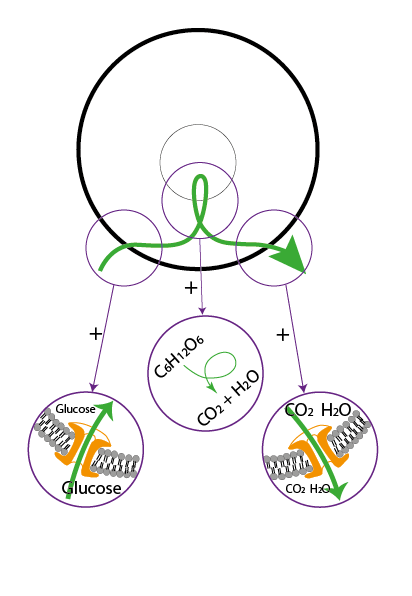
So, let’s face it: it seems that we were more or less asleep with our eyes wide open. Fact is that our society is now struggling in their attempt to minimalize the possible damage. The shift in time frames from country to country, from continent to continent, clearly shows how recipes and tactics are copied. Here and there some minor adjustments are made. And yes, apparently it seems to work. So, although we are copying, we do learn by experience from past and present and in doing so we learn to deal with this current situation.


And what do we osteopaths learn from this situation? Well, what we see and hear is that some are looking into their toolboxes, hoping to find an appropriate technique, while others dive into libraries in search for parchments from a distant past, hoping to find some wise instructions from the old doctor. … Seriously?


Has not Dr. Still already shown us his strategy and should not this be “common knowledge to all osteopaths”? And, isn’t it all about principles? Apparently not! At least it doesn’t look so when you read the questions and answers on the modern tool of communication: social media. The same tool that some modern osteopaths use to proclaim that Still’s principles, his philosophy is dated. Even more, some of them declare that it is just a relic from the past! “Science has evolved and so has osteopathy” – “hard” science is their justification. And in doing so, they justify the question: why bother teaching the past?


In these days, where patients are in distress, the consequences of this modern attitude become obvious. At this moment, a tennis elbow or painful back is not the topic of the day. No, this distress is of a different kind! So, this requires another tactic, another treatment, another technique! Is it? Is it really the case? … Because the basic principles of life are of all-times and by far no relics of the past. Nor do they belong to a specific dis-ease (A.T. Still, Philosophy of Osteopathy, page 2: It is my objective in this work to teach principles as I understand them, and not rules… knowledge for all diseases … .).


In the momentary raging storm that goes from country to country, from continent to continent, it seems that osteopathy has been put to the bench at the sideline. But keep in mind: in the coming weeks and months, we are all going to deal with numerous patients who will have “recovered” from more or less severe inflammatory processes. Clinically they have recovered, and it looks like that battle has been won. But they still carry the consequences. The question is how we are going to relief their dis-ease. So, how are we going to approach that battle? Let us try to resume Still’s “old dated” principles into a “young fancy” translation.

Where it begins


THE CELL


The first thing that we should ask ourselves is: where does it begin? Where does what begin? Simple: Life. By following the chronological steps, we start with energy, go to matter, continue to molecules, pass by the biomolecules and come to that complex adaptative system called “cell” (C. De Duve, Vital Dust – Life as a cosmic imperative, 1995; EVOST – Girardin-Höppner). The level of complexity, that is reached with the form of a cell, is connected to the term “life”. Many see this term as a noun. Instead it should be seen as a verb. It should be seen as a verb because it is a phenomenon. A phenomenon which can also be described as the behavior “being alive”. So, it is by far not just a stand-still-image.


BEING ALIVE


What should we understand by “being alive”? Well, it is nothing more and nothing less than a certain behavior that tends to maintain the original form. And whenever this is no longer possible, consequently that form will change. We can ask ourselves the question: where does this behavior “being alive” come from? To answer this, we must be aware that behavior is connected to the meaning of “form”. It means that “Form & Behavior” are one. So, if we want to understand behavior, we should also understand what form is about.


The cell has a specific form. And this results from a number of structural components and their positional relationships. Both characteristics play a significant role in the behavior called “being alive”. Both characteristics play a significant role in the tendency to maintain the original form although there are constant changes in the environment. Constant changes in the environment that put a strain on the structures and the different positional relationships between them. The structural components and their positional relationships are resisting that environmental influence (outside-inside phenomenon; A. Gurwitsch & H. Driesch, 1907, 1920, 1944).


Some authors describe the cell as a complex adaptive system. With other words: “complex” referring to a large number of relationships where the sum is more than the number of the parts (1+1=3). And, “adaptive” referring to the fact that the cell as a whole (a unity) is capable to respond to that environmental influence. This adaptive feature can be observed in two different ways. First, the cell is capable to respond without the immediate consequence that its form will change. Some authors refer to this as “the resilience of the form”. The most of us know this under the synonym of “metabolism”. Is this the only synonym? No, there exists a second possibility to observe the feature of adaption. In this case the resilience is not enough to resist the environmental influences. In this case the positional relationships between the structures of the cell cannot be maintained. The environmental influences can overcome that resistance. And as a consequence, the structuration of the cell will change – which means that the form changes. The adaption becomes a transformation, and this means either an apoptosis of the cell (decomplexification) or a replication by internal reorganization of that cell (complexification).


ABOUT METABOLISM AND FUNCTION


Every cell is characterized by a form. This form results from the structural components and their positional relationships. Both these characteristics allow the cell to maintain its original configuration. They allow the cell to maintain its form although there are constant changes in its neighboring environment. As we have just seen, this tendency to maintain the form can be described as the behavior of that cell. So far it is clear and simple. But.


The Babylonian confusion comes when we lose sight of that basic principle and begin to juggle with terms such as function.


Let it be clear: the basic behavior of a cell can be defined as “metabolism”. This metabolism can also be described as a function. Now where lies the difference? Simple. Let us describe the phenomenon of metabolism in detail.

To begin with: we have a cell and its environment. That environment is constantly changing. Whenever that changing environmental space also covers a part of the cell, the positional relationships between the structures of the cell come under strain. The behavior of the cell, the attempt to maintain its form, is now depending on different parameters: the first is the kind of environmental strain that is put on the cell. The second parameter is the configuration (structuration) of the cell itself. This means: any kind of behavior, any kind of metabolism can express itself in a different way depending on the variation of those two parameters. This different appearance of the cell’s basic behavior, called metabolism, will/can be described as a function. The way of describing it as a function depends on the contextual frame of the specific parameters.


So, let us look at the features of that metabolism. We should keep in mind that this is one continuous process. However, in most (if not all) books, this metabolism is split up in different steps of different single events posed in a linear fashion (illustration 1).


      • Step 1: something from the environment (example: sugar molecule) enters the space of the cell.
      • Step 2: the form of that same something becomes changed by the interaction with the “new” environment (the interior of the cell) – in physiology books this second step is described by numerous chemical reactions and interactions. These can happen immediately in that new environment (example: glycolysis) or only after another spatial change by entering a cell organelle (example= mitochondrion where the change of the form is described as Krebs cycle). Each single step in this chemical process is corresponding to the principle behavior, but only in different dimensions and levels of complexity! In physiology books these steps are all defined as functions. It’s a perfect demonstration of how something simple (principle) can be made incredible difficult (detailed example). Especially when the principle is not known or lost out of sight!
      • Step 3: the substrates (often defined as catabolites) of step 1 & 2 finally will leave the space of the cell and enter the environment. Without any doubt, each and every step of each individual catabolite can be described as a specific function as well.

But hey, why make it so difficult right at the beginning. Let us first understand the principle of “being alive”, let us first understand what metabolism is all about. (A.T. Still, Philosophy of Osteopathy, Chapter 10, page 168 ... our school is young, but the laws that govern life are as old as the ours of all ages).

Illustration 1.

Schematic drawing of a cell. The fat black circle represents the cell membrane. The circle almost in the center represents a cell organelle. In this case it could represent a mitochondrion. But it could also represent the cell nucleus or any other kind of organelle. The green arrow indicates the change in position of a molecule, in this case glucose. The small circles indicate enlargements showing what happens in that specific region. It corresponds with the three different steps that characterize metabolism.

(Source: JP. Höppner – Life as a verb, Maaseik 20?? coming soon)

IF ONLY WE WERE JUST ONE CELL!!


As long as we were just one cell, responding to our environment was not that difficult. Okay, starting with the moment of the “big bang”, it took about 10 billion years to reach the first form of a cell. But once we got there, it became quite easy to maintain that form. However, as soon as we became multicellular organisms, from that moment on, a lot changed.


In the beginning, the transformation of the cell went relatively slowly. It took about 1 billion years to change a prokaryote into an eukaryote. But once the eukaryote was formed, changes such as the transformation of a single cell into a multicellular organism went exponentially. It is interesting to observe that during embryological development these steps only take a few hours. Or a few days at the most. Presenting it here in writing even takes less time because the principle is quite easy.


Chronologically:

1) As long as metabolism is capable to answer the environmental stress, the form of the cell will not change.

2) Is this metabolism not capable of resisting this strain, the cell will change.

3) In the case of change there are two options: jumping down or jumping up in level of complexity.


The first means that the cell will fall apart into its separate structures while the latter means that the cell will multiply. Before this dimensional jump takes place, we can observe within the cell a phenomenon called “internal reorganization”. Looking closely, we can redefine a distinct chronology: metabolization  internal reorganization  complexification (multiplication) or decomplexification (apoptosis). This algorithm repeats itself all over again. In the end it will lead to a multicellular organism. (illustration 2)

Illustration 2

This picture shows chronologically the developmental steps of transformation based on a metabolism that does not succeed in maintaining the original form. The highest level of complexity based on the transformation resulting from metabolism is the cyst form. Although the level of complexity rises, in the end it is still a repetition of a simple algorithm: metabolization – reorganization – transformation.

The green arrows in the illustrations are referring to trajectorial metabolic movements (fluid dynamics).

(Source: JP. Höppner – Life as a verb, Maaseik 20?? coming soon)

Incorporating the environment


THE FEATURES OF A CYST


As soon as the form of a cell has been changed into a more complex organism, such as a cyst, the metabolic behavior of its cells will change. This is a direct consequence of the positional changes that takes place. As soon as the developmental stage of a cyst is established, the cells that are limiting the surface of the cyst are “caught” in between two different liquid environments. One on the outside and one on the inside. This is a completely new situation and it becomes obvious that this has an impact on the behavior of these cells: the cells at the surface of the cyst show a so-called “polarized metabolism”. This means that they receive their metabolic input from one side of the cell (step 1) whereas the metabolic output (step 3) happens at the other side.


Looking closely to the Form & Function of the cyst, we can distinguish an outer and an inner milieu. It seems a contradiction but in the case of a cyst, the inside (fluid chamber) is representing the outer milieu. This outer milieu is that part of space where the secretion will go to (the end of the metabolic story. The inner milieu, the so-called “source of the metabolic information”, is the real environment of the cyst. I know that this might be very confusing. But, in order to understand this one has to change its position by making a dimensional jump: look at the behavior of the cell and not to that of the cyst! This becomes very important as soon as we start to talk about tissues such as “frontier tissue” (also called “limiting tissue – see below). Often, we forget that the Form & Function of a tissue results from the Form & Function of its individual cells. In order to understand a tissue, we must make a dimensional jump to the cells and study their behavior!! In even more complex organisms such as the human body, the inner milieu is “the source of metabolic information”. So, although on the inside, the inner milieu represents the “metabolic” outside of cells and tissues! These basic landmarks as well as the basic behaviors are already established in this stage of existence.

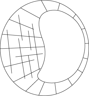
Illustration 3: the cells at the surface of the blastocyst are situated between two different liquid environments. The fluid chamber within the cyst represent the outer milieu because this is where the metabolic excretion goes to. This leads to an apparently contradiction because this means that the inner milieu is now on the outside of the cyst.

(see also illustration 4)

OUR BODY AS A CYST


Our body is a very complex organism. It contains many different compartments which are build up by many different tissues. Each and every single tissue is the result of multiple cell aggregations. The nomenclature is various and often quite confusing because different branches of science use their own terminology to describe the same Form & Function … However, it should not be that difficult. It is just a matter of understanding the principle.


Only two different kind of tissues? – Cells are the building stones of tissues. Tissues are those of organs and organs are the structural parts of an organism. Eventually all organs are the combination of derivatives of two different kind of tissues: frontier tissue an inner tissue. For each of them the different sciences have different terms.

    • Embryology
      • Frontier tissue – ectoderm & endoderm
      • Inner tissue – mesoderm
    • Histology
      • Frontier tissue – epithelium & mucosa (also neuronal tissue as well as endothelium and mesothelium in their behavior – both the latter however not in their origin)
      • Inner tissue – connective tissue (also muscle tissue)

(Please note that in some literature frontier tissue is also called limiting tissue – E. Blechschmidt)

(A strong support for this way of looking comes from oncology, there are only two types of cancers: carcinoma (frontier tissue) and sarcoma (inner tissue)

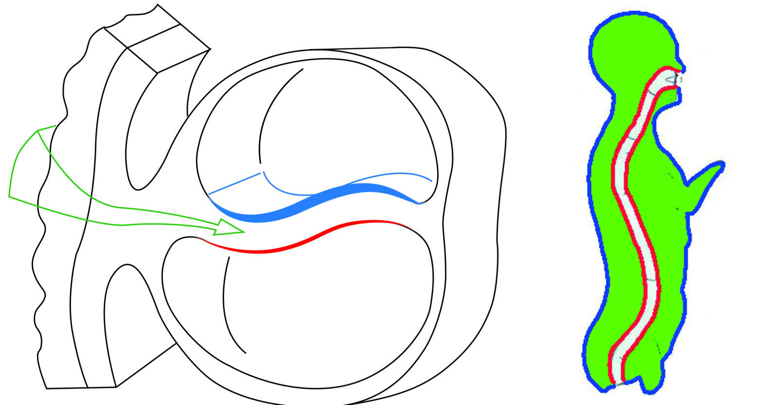
Illustration 4: In the left illustration we see the two germ layers ectoderm (blue) and endoderm (red). In between these two frontier tissues there is a small area called the inner tissue space. Within this space we can observe a fluid dynamic that is responsible for the metabolic behavior of the two germ layers (in later stage of development also for the mesodermal germ layer). In the illustration on the right we can see the presence of the two frontier tissues in a more anatomical shape. The large green space represents the location of the so-called connective tissue which is a direct derivative of the embryological inner tissue (mesoderm).

(Source: JP. Höppner – Life as a verb, Maaseik 20?? coming soon)

Frontier tissue – form & function – This tissue is characterized by the huge number of tied cells forming membranes. These cells and the membranes they form are limiting the border of the organism. You could say that the frontier tissue is the outer surface of the organism. The cells of this tissue are caught in between two different spaces, the inner and outer milieu. It is tempting to say that the outer milieu represents the environment of the organism. And in fact, it is. But only in this particular dimension. When we look into a different dimension, the dimension of tissues and cells, we notice that the so-called inner milieu is actually their environment.


How do we know this?

Easy, the space of the inner milieu provides the information that leads to the phenomenon of metabolism! This metabolism is polarized because the cells take up information on one side (inner milieu = environment) and excrete on the other side (outer milieu). The excretion of metabolic substrates creates a thin layer of secretion products at the outside of the frontier tissue, the outside of the organism. This layer of secretion substrates is partially the fertilizer or econiche of what is called the microbiome. The term microbiome is referring to a large number of organisms that live (partially) by the grace of the secretion products of the frontier tissue (illustration 5 & 6). The contemporary tendency of focusing and emphasizing the tiniest detail of this microbiome, regardless the fundamental meaning of the frontier tissue’s metabolic contribution, is a huge mistake! It is comparable to “microbiological pornography”: enlarging a tiny detail in such a way that it fills the complete picture in disregard to the totality and its unity in Form & Function. In fact, not always but all too often, it lacks an understanding of the principles behind Form & Function! – see later.

Illustration 5: schematic representation of the micro-organism of the skin

(Source: pinterest.com)

Illustration 6: schematic presentation of the micro-organism of the gut

(Source: http://www.trismegistos.lt/biologija/musu-nematomos-nuvertintos-ir-ignoruojamos-antrosios-puses/)

Inner tissue – form & function – This tissue is defined by the presence of 4 fundamental components: water, matrix, cells and fibers (chronologically in this order of developmental appearance).


The term matrix, in the context of this article, refers to all minerals and molecules, inorganic as well as organic, that are dissolved in water. Basically, every chemical form that does not look like H2O.

The number and variety of cells covers a wide range. All too often the number of them is so overwhelming that they hide the presence of the other components although these components have their meaning as well! (example: fat tissue - so many fat cells that all other components become overshadowed although they are present; in the dissection room many participants see so many yellow fat cells that they do not even recognize the fibers of the connective tissue, in fact, they do not even see the whole as a form of connective tissue!)

When it comes to fibers we can distinguish to basic types: collagen and elastin fibers.


In the earliest stages of development, the inner tissue is characterized by the presence of mainly water and matrix. Only a few cells are present. In most textbooks these cells are called mesoderm cells. In most textbooks it is even simply defined as mesoderm, leaving out the fact that the cells of this germ layer are completely surrounded by water and matrix!


All too often this leads to a mental/intellectual disconnection of the importance of these components. For instance, the importance for the behavior of these cells!

During the ongoing transformations of the inner tissue, the number of components as well as their variety and distribution changes. Consequently, we can observe future connective tissue in many different forms and behaviors. One most extreme represents the most fluid form: connective tissue in a liquid state such as blood. The opposite extreme represents the densest form: connective tissue in a solid state such as bone. In between these two extremes the variety goes from loose connective tissue to ligament, fascia’s, tendons, etc.. And each and every single one has a different behavior, a different function. In fact, we can distinguish two basic behaviors of connective tissue: a chemical and a physical behavior. The chemical behavior of connective tissue refers to the state of permeability. The state that allows an exchange of chemical information and determines the metabolic activity in that space. The physical behavior refers to the resistance of the connective tissue to a physical strain. It should be emphasized that the more a connective tissue tends to high permeability, the less resistance it can offer – and vice versa. Observing both behaviors we come to the conclusion that “being alive” is a constant search for compromise between the two basic behaviors of connective tissue. And this leads to a specific form that comes in the same large variety as its compromises. (Girardin, Osteoporosis mischaracterisations, 2019))(illustration 7)

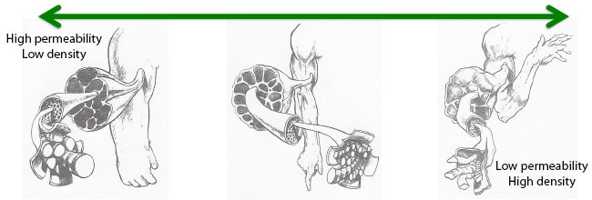
Illustration 7: this illustration shows the scale of permeability and density in relation to form of the locomotor system.

(Source: S. Keleman, Emotional anatomy, 1985)

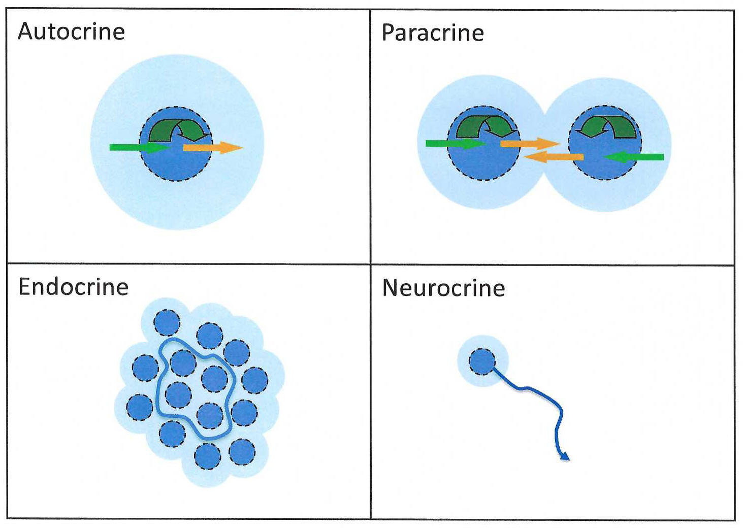
ABOUT COMMUNICATION SYSTEMS


Epithelium and mucosa, both frontier tissues, are characterized by a polarized metabolic behavior. They receive their metabolic information from the environment called connective tissue (inner tissue) and secrete their “catabolites” into the outer world. So, the metabolic activity of the frontier tissue derivatives is directly connected to the condition of the inner tissue. In fact, what we can say is that the metabolic behavior of the frontier tissue is determined by the condition of the connective tissue, chemically and physically. Both conditions determine the transfer of metabolic information. In textbooks of physiology, this transfer (and its consequences) is described in the chapter of the so-called “communication systems”. We can distinguish 4 different communication systems if we want to box them in a drawer:

Autocrine – Studying this communication system shows us that the metabolism of a single cell finally leads to a self-regulation of its own behavior. Meaning, as soon as the “original” chemical stimulus is metabolized, the activity slows down. Please keep in mind that physical stimuli can and will have an impact on the cell’s behavior as well.


Paracrine – In this case the metabolism of two neighboring cells influence each other. This means that the substrates produced by the metabolism of one cell will represent a stimulus for the metabolism of its neighbor. It shows us that the meaning of anabolite and catabolite should be seen as quite relative. After all, the catabolite secreted by the metabolism of a certain cell becomes the anabolite for the neighbor.


Endocrine – This communicating system is also known as a “telecrine system”. The latter simply refers to the fact that the communication takes place over a longer distance. It means that, unlike in paracrine conditions, not so much “only” the neighboring cell will become influenced, but much more those in a far distance. In the case of an endocrine communication system, the term often leads to the reflex of translating and reducing this behavior to a single term called “hormone”. Please keep in mind that in its basics not the substrate, a specific hormone, should receive the attention but initially much more the transport system for this communication! Not that the hormone has no meaning, but keep in mind, when the substrate is not transported into a certain region, the effect on the metabolism of the other cell will be …(?). Exactly: neglectable! In the case of the endocrine communication system the transport is assured by the vascular system, in particular the veno-lymphatic system! A full understanding of the landmarks regarding this communication system is a premise to understand the metabolic behavior of a complex organ such as the human body. With other words: learn the anatomy of the venous and lymph vessels!!


Neurocrine – It is also a telecrine communication system. But in this case not the vascular system is responsible for the transport of the metabolic substrate but the cell itself. Or at least a part of the cell is responsible: the extension called axon and dendrite. When we look closer into the exchange of metabolic information, the neurocrine communication is, just as the endocrine, a paracrine phenomenon. In the context of the neurocrine regulation of metabolism, we have to emphasize on the autonomous nervous system. Here we distinguish two different systems: the parasympathetic nervous system and the (ortho)sympathetic nervous system. One of the main differences is the fact that the parasympathetic nervous system is communicating with the direct neighborhood (inner tissue space) of the frontier tissue cells – the neuron spits directly into the frontier cells neck – whereas the (ortho)sympathetic nervous systems communicates with the frontier tissue cells by influencing the larger vascularization of their neighborhood. (for instance by regulating tonus of precapillary sphincter = opening and closing the gates).


Cleaning up the room, cleaning up the house


Studying the chapter “immunity” all too often leads to a study of schemes that contain a large variety and number of names related to cells and molecules. It makes many heads spin and, in the end, we get lost it the woods of incredible complicated “metabolic” relationships. Studying the chapter “immunity” all too often leads to the fact that the basic landmarks and trenches get out of sight and this leads to a battle of the apparently blind who is almost looking desperate in the attempt of trying to kill the enemy. An enemy that seems to lurk behind every tree or bush. Can we please hold our horses and look at the basic principles before we plunge into the details – please?


(RE)DEFINING IMMUNITY


What is “immunity”? In fact, it is not so difficult to understand. At the end of the second week during embryological development, with the appearance of the axial process (also known as notochord or chorda dorsalis), cells are added to the inner tissue space. This space is located between the frontier tissues ectoderm and endoderm. At this very moment the future embryo is receiving a new kind of cell, a cell that has no polarized metabolism. This means that these so-called mesodermal cells spit their substrates into their own neighborhood and into the nearby space of the frontier tissue cells. This has consequences.


The first consequence: messenger – The first consequence is that the metabolism of these inner tissue cells alters the composition of the environment of all other cells – neighboring inner tissue cells as well as the cells of the epithelium and mucosa (frontier tissue cells). In this context we have to keep in mind that the frontier tissue cells receive their metabolic information by the backdoor which is located in the inner tissue space (importance of basal membrane)!! Consequently, the behavior of the epithelium and mucosa is directly related to the condition of that back-door-space, chemically and physically!


The second consequence: cleaning part 1 – The second consequence is the fact that the cells of the epithelium and mucosa are metabolizing information coming from the inner tissue space. Due to their polarized metabolism we could say that by the metabolic activity of frontier tissue cells, the inner tissue space becomes cleaned. The frontier tissue cells therefor also clean up the mess produced by the inner tissue cells. In diagnosis this is all too often forgotten: the condition of the secretion layer that covers the epithelium and mucosa is a good indicator for the metabolic activity of the frontier tissue and the condition of the inner tissue space (how clean or dirty is the garbage can)!


The third consequence: cleaning part 2 – The third consequence is that the metabolic activity of the inner tissue cells can be seen as a cleaning activity as well. Some of the cells of the future connective tissue become “specialized” in metabolizing specific agents. These “disturbing” agents come in different forms such as molecules (small and large) and even cells which become recognized as “aliens”. As soon as some inner tissue cells start to clean up the mess, this specific kind of metabolic behavior can be defined as immunity.

In a worst-case scenario, the inner tissue cells even start to “digest/clean” cells of the body itself. In that case we speak of autoimmunity.

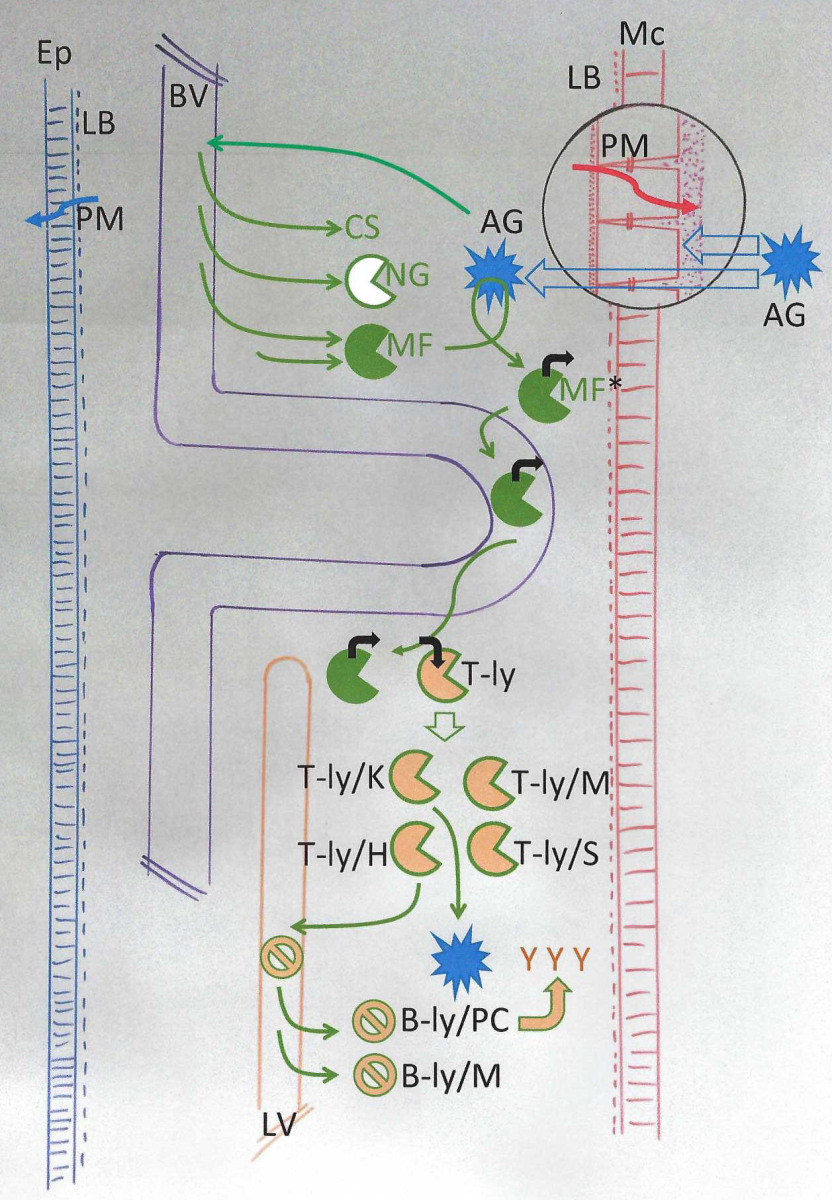
PUTTING TRENCHES INTO PERSPEKTIVE


(Illustration 8: schematic representation of the different trenches in which metabolic activity takes place under the title of immunity.)


The first trench: surface of the frontier tissue – In the outer world, there are a large number of “Antigen (AG)” that can be seen as aggressors. They come as bacteria, viruses, parasites etc.. When these agents are attacking the human form, they are confronted with several trenches. It is comparable with a hostile army in front of the city walls. The walls of the city are built by the cells of the “Epithelium (Ep)” and “Mucosa (Mc)”. The cells of these tissues are standing on a firm fundament called “Lamina basalis (LB)”. This basal membrane not only keeps the cells in their proper position, the basal membrane also contributes to the polarized metabolism (PM) of this tissue. Epithelium and mucosa, these frontier tissues are like walls that are difficult to breach due to the fact that the bricks are closely packed together. This is especially the case when it comes to the epithelium of the skin (blue color). In the case of the red wall there are minor gaps between the cells. This might explain why an attack from the outside could be more successful when trying to break through the red trench. Nevertheless, in both cases it is difficult to get through. Not only because the frontier tissues represent a physical trench but also a chemical one. Because both cell layers produce secretion products (PM, polarized metabolism) and these substrates are covering the surface of the organism. These products can be compared with a swap, the wetland that surrounds the city. It makes it difficult to cross. An additional help to that defense is the fact that we can observe a large number of mercenaries who benefit from that specific environment (microbiome). In return they work for us in defense. However, although they are large in number, they are not specific in the job they do (which is metabolism!). And if poorly fed by the frontier tissue cells, their number might be poor or might not have enough strength to continue a longer combat. (see later)

The second trench: the inner tissue space – Once the hostile perceived antigen is getting into the city, after passing through the wetland and breaching through the walls, they enter the so-called inner tissue space. This is the next line of defense, the next trench. Present are water, matrix, cells and fibers. All of them contribute specifically to a defense in the case of a possible breach of the walls. The matrix component is referred to as the “complement system (CS)”. The concentration of that matrix component will increase. This elevation in concentration happens as soon as certain chemicals (such as histamine and bradykinin) cause a vasodilatation in the blood vessels (BV) and change the permeability of these vessels (part of the phenomenon called inflammation). This complement system consists of chemicals (proteins) which will attack some of the antigen (for instance membrane of bacteria) and also marks the antigen. This marking of the antigen leads to attraction of leukocytes belonging to the mononuclear phagocyte system. This is the cellular component of the inner tissue space that also contributes to the defense.

The cells of the mononuclear phagocyte system come in different types. The first cell that the antigen encounters is the “Macrophage (MF)” which, just like the complement system, belongs to the a-specific defense. Some of these macrophages are already present in the inner tissue space and are defined as histiocytes. Their numbers will become enforced as soon as “Neutrophil Granulocytes (NG)” leave the vascular system and join the battle (the NG also belong to the a-specific defense). Shortly after them new macrophages will arrive in the region of the attack. As long as these “new” macrophage are still in the vascular system, they are called monocyte. (I know, why make it so difficult, when I leave the house, I do not change my name either!). So as soon as the monocyte becomes a macrophage by leaving the capillaries, they too will start to metabolize the antigen. An important difference with the neutrophil cells is the fact that this macrophage lives longer. Consequently, it can attack the antigen for a longer period of time and, also important, it can clean up for a longer period of time.


The third trench: the veno-lymphatic system – Although this trench belongs to the inner tissue space as well, it requires a special attention. A macrophage who has successfully attacked and conquered an antigen can and will re-enter the vascular system. In doing so it is capable of meeting a specific cell called “T-lymphocyte (T-ly)”. The macrophage has a chemical sign on its membrane. When this sign fits a receptor of the T-lymphocyte, this will activate the metabolism of the T-cell (paracrine communication). Consequently, 4 new cell type will originate from this metabolic T-cell behavior. The cell that is called “T-lymphatic memory (T-ly/M) is the one that will remember the antigen for the future. The “T-lymphatic killer (T-ly/K)” is the one that will attack the antigen as soon as possible whereas the “T-lymphatic suppressor (T-ly/S)” will assure that the attack does not go out of control. The fourth and final cell is the “T-lymphatic helper (T-ly/H)” and this one will activate the so-called B-cells.

The B-cells are also, like the T-cells, belonging to the specific defense system. These B-cells are located in the lymph nodes (LV, Lymphatic vessel). Once they become activated (paracrine communication), we can observe two metabolic responses. The first response is that they will produce memory cells (B-ly/M). the second response is that the so-called “Plasma cells (B-ly/PC)” will produce antibodies that will attack the antigen. These anti bodies are called IgA, IgD, IgE, IgG and IgM. These molecules will connect to the antigen and function as a marker that makes it easier for a macrophage and T-killer to find the antigen. The antibody will also neutralize the toxic substances and activate an immune complex (Antigen-Antibody-Complex System).


The assistant cleaning manager – preparing the trenches


Every organism is under constant influence of the environment. The human body is no exception to that rule. Every organism is responding to that influence. And apparently it works quite well. After all, the human body is the result of an evolutionary try-and-error of 13,5 billion years since Big Bang. So how come that from time to time we have the impression that this “defense mechanism”, this “cleaning mechanism” apparently does not seem to function as it should be? There are several possibilities.


Each form is a consequence of its structures and their positional relationships. These structures and their relationships are responsible for the resistance which the form can bring up against a strain from the environment. An adequate response to that environmental influence depends on the premise that all of these structures are in the “right position”. We can define this state of the form as being “the normal” (A.T. Still). As soon as we are dealing with “the abnormal”, the form is up to a certain extend “crippled” in its response of resistance. Consequently, the more we are dealing with a situation where relationships have been changed, the less adequately we can counter these environmental influences. We have less resilience.

But this is not the only possibility to explain why our defense/cleaning mechanism does not behave properly as it could. Let us just assume that our form is not crippled by changed relationships between structures. With other words, the situation in which we could say that our body is in a healthy state. In this state our form is capable of dealing with the environmental stress with only little effort. Now it is merely a matter of amount of stress and period of time in which the stress is present. With other words, two questions important: A) is our body already preloaded by stress combined with an additional stress that we have to deal with and/or B) is the amount of stress simply too much to handle for any healthy body at such short notice? Depending on the answer to these questions the therapeutic procedure will be determined. So, how can we prepare the trenches for the coming battle?


CLEANING UP THE GARBAGE


By knowing the landmarks, we become aware of the fact that the wetland that surrounds our body plays an important role in the first line of defense. So yes, the microbiome should not be ignored. But some smoothies or other healthy potions will not do the job. Even if we add some extra-soldiers in form of probiotics, we should keep in mind that this can have consequences in the opposite direction of what we intended to do (Hempel et al 2012, Bafeta et al 2018, Rao et al 2018, Lerner et al 2019).


So, this is too simple and can even backfire.

With other words, if we want a good swamp that surrounds us in order to make it difficult for the attacker, we need a solid metabolism that provides it! We need a good secretion of appropriate substances that make the microbiome flourish in such a way that it can play its role of first defense. And this means that we have to assure a good condition of the inner tissue space! And this we do by assuring that the garbage within the inner tissue trenches is cleaned up and that these inner tissue trenches are ready to act. To do this we need to put the players in position.


PUT THE PLAYERS IN POSITION


Let’s get physical – We have seen previously that the connective tissue represents two different features. On the one hand we have a chemical and on the other hand a physical feature. Although both have their own characteristics, we can and should not deny the fact that these characteristics are bound to each other. For instance, we cannot deny the fact that a physical strain on our body changes the permeability of our inner tissue space. The same space where the chemical exchanges takes place. The same space where metabolic events take place. Increase of the tension in the fibers of the connective tissue changes the viscosity of the fluid immediately. Increasing tension in fibers leads to increasing fluid viscosity and this leads to decreasing permeability and consequently decreasing exchange. This affects the metabolic behavior. This leads us to the conclusion that in order to optimize metabolic exchange, in order to optimize our “cleaning defense mechanism” we require a body without strain! (A.T. Still !!).


No recipes, but principles – It is all too easy to use a recipe for all patients who are dealing with symptoms related to immunity. In fact, it is all too easy for any kind of symptom. But, instead of recipes, here are some principles to keep in mind:


  • Nourishment: it should be obvious that if you are eating and drinking like a pig, in due time your connective tissue will look like a gigantic garbage bag. And who will have to do the cleaning? Correct, the cells of the inner tissue which belong to the immune system. So, having an eye for the diet of the patient might be a good idea. The question now is: what’s a good diet? This is a hot topic because nowadays it seems that food has become the subject of a new religion! And many new gurus are spitting quite a lot of information. But let’s be honest, quite a lot of them don’t even know the meaning of the word physiology and have no idea of the consequences of their so-called healthy diet. It is okay to have an opinion that does not comply with the common mainstream, but certain aspects of chemical events in our body are a certainty. Just like gravity, we cannot deny that it exists. A golden rule: keep it simple and keep it various. (Note: some additional nutrient can be added in order to support some chemical reactions – see later)


  • The cleaning staff: it speaks for itself that, when it comes to excretion in order to get rid of the chemicals in our body, the epithelium as well as the mucosa are the biggest in numbers (we should say in surface). But, their metabolism, their excretion behavior, depends on a number of control mechanisms. And these control mechanisms can suffer from physical strain. So, here is a check-up list:
    • Autocrine/Paracrine: tensional strain in the connective tissue has a repercussion on the exchange possibilities. The tensile stress can and will have an effect on the permeability in the region where the strain occurs. This region can be very local as well as covering a larger area. In general, these tensional strains are characterized by a specific direction. The general idea is to get rid of this tensile direction. Which technique should be used and in what direction we should go cannot be predicted neither can it be generalized. Keep it simple by using the principle: A healthy state is a state of no pattern.
    • Endocrine: when it comes to the endocrine communication system, we have to distinguish two aspects: the venous and the lymphatic aspect.
      • VENOUS – a profound anatomical knowledge of the venous vascularization is a premise to be able to work on this communication system. The capillary system is an essential piece of the puzzle when it comes to guaranteeing an adequate condition for metabolism – for providing as well as for disposing. Pattern of strain that cross the direction of the flow in these capillary structures tend to cause congestion. And this is something we do not wish to have (congestion is one of the characteristics of inflammation!). So, the previous principle is once more valid: a healthy state is a state of no (congesting) pattern.
      • LYMPHATIC – regarding the lymphatics we are dealing not only with larger vessels such as the lymphatic duct or the large number of lymph nodes. In relationship to the immunity aspect we should keep an eye on the importance of the so-called MALT system. MALT stands for ‘”Mucous Associated Lymphatic Tissue”. In fact, over the years this term was extended by other terms such as GALT (G = gut), and SALT (S = skin) and so on. Over the years it became obvious that this lymphatic association belongs to any kind of frontier tissue derivative (a teaser: glymphatic system of the brain). The lymphatic part of the endocrine system is part of the connective tissue. Congestion caused by directional strain should be avoided! And please keep in mind, it is not because the patient is coughing, that a strain in the abdominal digestive system is of no importance for the patient’s throat symptom!!
    • Neurocrine: as mentioned earlier, when it comes to the neurocrine communication system, the autonomous nervous system plays an important role. Please keep in mind that any kind of strain that runs spiral-diagonal or even transversal to the trajectory of a nerve extension (axon and dendrite) can and will cause a disturbance for the axoplasmatic transport within that nerve. And consequently, the nerve will do not his job as it should be! Getting rid of a perpendicular pattern caused by a transversal structure, such as a diaphragm, is a must.


Important landmarks – From an osteopathic point of view certain anatomical landmarks are important in order to keep the trenches in good shape. Here some of the most important ones:


  • The cleaning areas: when it comes to anatomical regions that we should keep an eye on, here is a short list:
    • A longitudinal space: this space represents the anatomical regions of the cervical spine, the mediastinum, the retroperitoneal space as well as the pelvic region (illustration 9). Interesting to notice is that the complete veno-lymphatic as well as autonomous nervous system is present in this inner tissue space. It is easy to imagine that any kind of strain, and especially in form of a transversal pattern, will cause a disturbing influence on the functionality of these structures! This should be avoided at all times and at all costs!! A profound anatomical knowledge of all these longitudinal orientated structures and those who cross them is a significant premise for an adequate treatment!!
    • A transversal space: referring to this transversal space is referring to all transversal structures in the human anatomy. In fact, we could list them under a common title such as “diaphragms”. But keep in mind that “classically” we only distinguish the larger ones such as the Tentorium cerebelli, the mandibular floor and hyoid, the upper thoracic opening (Apertura thoracis superior), the abdominal diaphragms (3 portions, each with distinct relationships!) and finally the diaphragm of the pelvic floor. But, in reality any structure running transversally to neuro-vascular highways should require our attention. Structures such as ribs but also less dense ones such as for instance the mesocolon transversum or mesenterium.

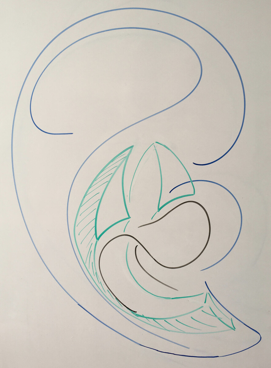
Illustration 9: schematic presentation of the inner tissue space (green) that extends from the base of the cranium till the end of the pelvic region. This space develops as the abdominal diaphragm (black) becomes repositioned. This repositioning is a complex movement – peeling away from the spine (spinal cord & brain), horizontalization (liver & hearth influence), relative descending (digestive system). The diaphragm represents a transversal structure can influence the longitudinal organized inner tissue space and al of the blood vessels and nerves that is contains.

(Source: Anatomie & Ontogenesis – seminar 4: Homunculus interni ; Wroclaw 2019)

Tagline

  • Connective tissue:
    • In general: we can look at the connective tissue as one gigantic potential battlefield. Earlier we have emphasized that this connective tissue has two important behaviors, a chemical and a physical one. It should be clear by now that the chemical behavior is directly related to the cleaning and defense action of the connective tissue. As osteopaths it should be our focus to optimize this metabolic action by assuring the absence of physical strain. Physical strain hinders the permeability and consequently the metabolic actions.
    • More specific: Treatment of fascia, aponeurosis, ligaments etc. is one level of complexity (those of the fibers) that we should keep in mind. Especially because these structures have specific characteristics when it comes to their fibers. These fibers, once under strain, expel fluid and densify the connective tissue. But also, these fibers and their directional characteristics can and will have an influence on neuro-vascular structures. However, connective tissue also consists of more than just fibers and this means that we can also work physically for instance on the fluid component (features of a fluid current!).


  • Vascular system – venous part: when it comes to the venous system, we have to keep the following in mind: we have three (almost) separate venous systems – a portal, a caval and a vertebral venous system (Herlihy 1947, Batson 1956).
    • In general: These systems communicate in specific areas with each other and this means that especially these regions are important (such as the region of the Fossa pterygopalatinum where the Plexus pterygoideus communicates with the Sinus cavernosus – just to mention one of the various communicating areas). These regions are so important because when a strain hinders the communicating flow here, it can cause congestion in one or more of the venous parts! So, keep an eye on these anatomical regions (all too often we know them as diaphragms). Beside these communicating regions, there is also the flow in each system itself. This flow should be guaranteed at all times. See here the details:
    • The vertebral system: For the vertebral system this means that a strain which can hinder the flow in the vertebral column should be treated. Please do not forget that these vertebral veins are in continuity with the dural sinuses! This becomes even more obvious by the anatomical relationship with the Dura mater. See also the veno-lymphatic pump mechanism (Guy 1930, Batson 1956, Girardin 1996, Kraml 2013).
    • The portal system: For the portal system the relationship with the liver is a very important landmark, for various reasons. To understand this, we have to keep in mind that the so-called liver is nothing else than the combination of two distinctly different organs: a vascular organ and a metabolic organ (exocrine gland). The vascular organ consists of the large number of capillary blood vessels (liver sinuses). The metabolic organ is represented by the gall system (hepatocytes are in fact nothing else than the mucosa cells of the gall ducts!). Both organs communicate by the inner tissue space called “space of Disse”. In this region of the liver not only the blood flow is regulated, but also the excretion of substances. The liver as one organ behaves as a kind of filter. This filter does not only regulate the viscosity of the blood but also cleans the inner tissue space (Disse)! Congestion in this region will hinder the effectivity of the immune system – locally due to the influence on cleaning cells within Disse (Kupffer cell, Natural Killers, Large Granular lymphocytes) but also systemically because it will influence the condition of the inner tissue in other regions as well. This congestion can be the consequence of two different origins: 1) either the vascular system is hindered by physical strain coming from structures that influence the flow. Structures such as for instance the mesocolon transversum, mesenterium and mesosigmoideum. But also smaller structures such as Lig. pancreaticolienale can cause (local) congestions (ligaments of peritoneal origin!); 2) or it is a question of chemical overload related to increased metabolic activity (endogenous) or digestion (exogenous). In this case we can use besides a physical also a chemical approach by adding specific substances that support bile production as well as its excretion. In a worst-case scenario, both are present, physical and chemical.
    • The caval system: This part of the venous system can be classified to the locomotor system and to the urogenital system. Consequently, it leads to the conclusion that for instance strain within the locomotor system can and will have an impact on the flow within the Vena cava. Transversal structures such as the upper thoracic opening (Apertura thoracis superior) can represent an important strain on the flow within this venous system. In fact, any kind of diaphragm, any kind of transversal structure that stands more or less perpendicular to the flow can cause congestion. Or at least it can cause a slowdown of the flow and consequently the metabolic activity within the region related to that blood vessel. But not only larger structures such as diaphragms can cause this phenomenon. Smaller structures such as ribs or even tiny ligaments can be a cause. And last but not least we should not forget that the veins belonging to the cava system are situated within a special place. They are located within that longitudinal space that extends from the base of the cranium into the mediastinum, from their continues into the retroperitoneal space to end up in the minor pelvic region (see illustration 9). Within this extensive space the venous systems vertebral-portal-caval “communicate” with each other! Any kind of strain coming from a transversal hindering structure will put a burden on the cleaning staff in the trenches.
  • Vascular system – lymphatic part: related to this landmark we can conclude that the remarks and attentions discussed in relationship to the venous system are valid here as well. Of course, there are special additional landmarks related to the lymphatic system, for instance the location of the lymph nodes, but in general most of the hindering structures for venous flow will also have consequences for the lymph flow. We should once more emphasize here the special location of many main lymph structures within that space that extends from the base of the cranium till the pelvic region! So pay attention to special locations such as for instance the “ring of Waldeyer” in the cervical region or the “plaques of Peyer” in the iliocaecal region of the gut.Supplementary, treatment of the pleural and peritoneal relationships should also be considered whenever necessary – see previous remarks on MALT etc.. (illustration 10)

Illustration 10: Short theoretical introduction and overview of important landmarks in the visceral system related to the vascular system before participants continue their dissection assignments.

(Source: Heidelberg Dissection seminar February 2020)

Conclusion


What is the recipe to deal with inflammation and even infection? In its basic it is simple:

  • Be gentle for your garbage can
    • Avoid filling the bucket up to its edge
    • Empty the bucket (in time)
    • Assure that the cleaning staff can do its job


How do we do it?

  • Know your landmarks by studying anatomy
    • This helps you to check on the condition of your trenches
    • This helps you to recognize where the hindering strain is


What tool(s) do we use?

  • For osteopaths mainly physical tools – manual work, adequately determined by the knowledge of:
    • Every landmark has a specific form
    • Every form is resulting from structural components
    • Every positional relationship requires a specific approach
      • Physically but also mentally (visualization!)
    • Chemical support can be used – catalyzation of chemical reactions
Book mockup

Discover the book

Life as a verb